The raw signal
Injected ICG fluoresces for a short window. On the endoscope feed the signal is diffuse, and it is read by eye.
 Contact
Transforming fluorescence into real-time anatomical and functional insight during surgery.
Injected ICG fluoresces for a short window. On the endoscope feed the signal is diffuse, and it is read by eye.
Time-series analysis of the same feed separates vessels from surrounding tissue by how the signal arrives and clears.
The map is drawn on a second monitor. No new instruments, no new steps, no change to the sterile field.
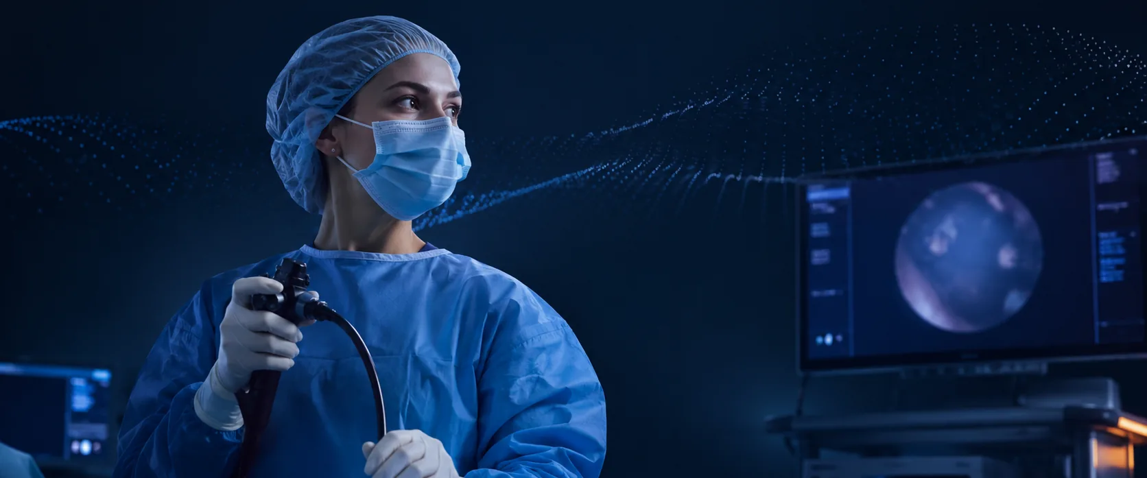
Fluorescence-guided surgery is moving from a niche imaging technique to a routine part of the surgical workflow. The way the signal is interpreted has barely changed.
Minimally invasive and robotic approaches increasingly make the camera the surgeon's primary view of the anatomy. That makes the information carried in the image more important — and it makes software interpretation possible.
Near-infrared imaging is increasingly built into surgical platforms, and ICG is already used across multiple procedures. The hardware and the signal are entering the operating room at scale.
Fluorescence is dynamic, spatial and time-dependent, but most systems still present it as a transient green image. The underlying signal holds far more information than a surgeon can extract visually in real time.
The software under development converts a near-infrared video feed into a structured map of the vasculature. It analyses how the fluorescence signal changes across frames rather than processing each frame on its own.
Segments and classifies tissue in the field, separating vascular structures from parenchyma.
Tracks that tissue through breathing, retraction and camera movement, so the map stays on the anatomy.
Differentiates arteries from veins from the temporal phase of the signal — the order in which vessels fill and clear.
Camera-agnostic. It works alongside the fluorescence systems already in the theatre — Stryker, Karl Storz, Olympus, Intuitive.
On device. Processing runs locally. There is no cloud dependency during surgery.
Workflow-neutral. No new steps in the sterile field, and nothing new for the surgeon to hold.
The surgeon decides. The system presents a reading. The clinical decision stays with the surgeon.
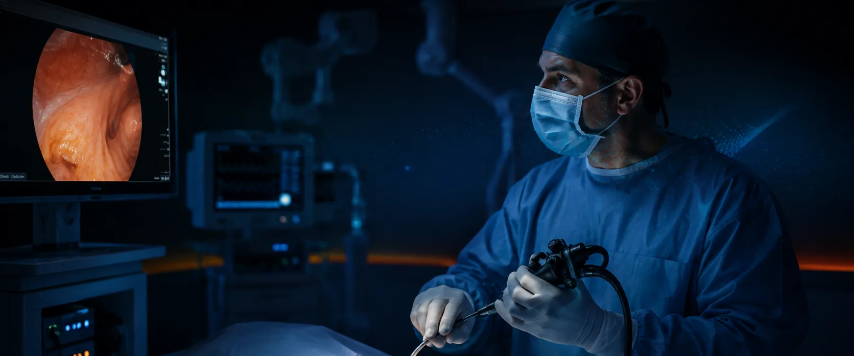
Development happens in the theatre or it does not happen. Collaborations with leading Danish cardiothoracic centres are being formalised, and we have room for more.
Two lines on your department and your case volume is enough to start.
contact@navisurgvision.comNavisurg Vision continues a body of work on reading fluorescence signal quantitatively rather than by eye, carried from perfusion imaging into thoracic surgery.

Former VP of Software Development at Perfusion Tech, where he led PerfusionWorks, a CE-marked fluorescence-guided surgery platform. Former Director of Software Development at 3Shape. Leads strategy, clinical and regulatory.

Former software architect at Perfusion Tech, Aiia and 3Shape. Leads software architecture: real-time image processing, embedded systems and medical software.

Surgical imaging, algorithms, GPU and AI. Ex-Perfusion Tech; full-time on the vessel-visualisation core.

Serial entrepreneur, 20+ years in Danish medtech and biotech. Fifteen-plus exits and two IPOs, among them ChemoMetec, SynAct Pharma and CLC Bio.

Co-founder and CSO of Perfusion Tech. MD, University of Copenhagen, and co-inventor of the core IP.

Co-founder and CEO of Perfusion Tech. DTU engineer, and co-inventor of the core IP.

CMO of Perfusion Tech. Twenty-plus years in biotech and pharma.
Thoracic surgery clinical anchor. Professor emeritus, engaged as an external consultant.
Surgeons, research units, and people who want to build this: the same address reaches us.
contact@navisurgvision.com